Anatomy Of Chest And Lungs - Amazon Com Male Chest Anatomy Of Thorax With Heart Veins Arteries And Lungs Poster Print By Leonello Calvettistocktrek Images 34 X 22 Posters Prints / Both tlc and lung compliance are measured for the diagnosis and treatment of various lung disorders.
Anatomy Of Chest And Lungs - Amazon Com Male Chest Anatomy Of Thorax With Heart Veins Arteries And Lungs Poster Print By Leonello Calvettistocktrek Images 34 X 22 Posters Prints / Both tlc and lung compliance are measured for the diagnosis and treatment of various lung disorders.. The base is the concave lower surface of the lung that rests over the diaphragm 9. Aug 09, 2019 · chest bone, ribs, lung, heart, xiphoid process, sternum anatomy. A serious lung condition characterized by scarring, inflammation, and narrowing of bronchioles, the smallest of the airways. See full list on therespiratorysystem.com The structures in the lungs directly responsible for the function of respiration collectively form the lung parenchyma.
The right lung has a deeper concave base as it is positioned higher to make space for the liver located beneath 7, 30. The left and right lungs are situated on the two sides of the body with the heart, another vital organ in the thoracic cavity, located a little in front of, and at the middle of them 5. The inferior mediastinum is larger between the two and further separated into the posterior, middle, anterior and mediastinum 23. You will also find the xiphoid process, 10th rib, the apex of the heart, the coronary vein of the heart, cardiac notch of the left lung, 4th rib, sternum, manubrium, ıst rib, the apex of left lung as well. This root not only connects the two lungs with each other but also keeps them suspended in the thoracic cavity.
Lung Anatomy Physiopedia from i.ytimg.com Structures including the bronchus, bronchial veins and arteries, pulmonary artery, two of the pulmonary veins, pulmonary plexus of nerves (anterior and posterior), and lymphatic vessels bundle together to form the root of the lungs 9, 10. The surface between the left and right lungs, it houses the hilum. Mar 18, 2015 · the chest is the area of origin for many of the body's systems as it houses organs such as the heart, esophagus, trachea, lungs, and thoracic diaphragm. Some of these get trapped in the nasal cavity by nose hairs to cleanse the air partially. Both bronchi and bronchioles are encircled with hyaline cartilage rings to help them maintain their shape 34. You will also find the xiphoid process, 10th rib, the apex of the heart, the coronary vein of the heart, cardiac notch of the left lung, 4th rib, sternum, manubrium, ıst rib, the apex of left lung as well. They are of different sizes and are divided into multiple lobes 3. The nerve supply to the lungs and some of its associated organs and muscles comes from branches of the vagus nerve and the phrenic nerve 7.
The space is divided into the inferior and superior mediastinum.
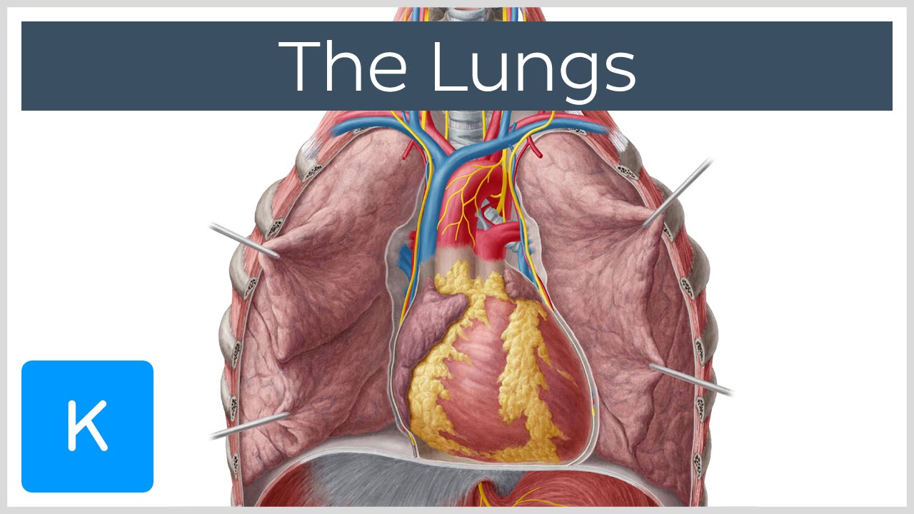
Both tlc and lung compliance are measured for the diagnosis and treatment of various lung disorders. As the name suggests, this is a concave basal surface, facing the diaphragm. It is the sum of the air released by the lung after a maximum exhalation (vital capacity or vc) and the volume of air left behind within the lungs after a deepest exhalation (residual volume or rv) 46. The two layers form a hollow space between themselves, which is known as the pleural cavity and is filled with pleural fluid, so the two pleural layers do not stick to each other 21. It is often incurable, most cases being managed with medication that prevents further blockage of the bronchioles. On the inferior surface, the lungs are bordered by the diaphragm. This deoxygenated blood is then pumped back to the lungs by the right ventricle via the pulmonary artery. See full list on therespiratorysystem.com Even though there are only two lobes, the upper lobe has a projection, referred to as the lingula (meaning little tongue in latin) that serves as an equivalent to the middle lobe of the right lung 15. Some of these get trapped in the nasal cavity by nose hairs to cleanse the air partially. Then, the cells receive the oxygen and send the carbon dioxide produced by the cellular functions into the blood to be carried by the superior and inferior vena cava veins to the right atrium, from where it reaches the right ventricle. When we inhale, the air enters through the nasal cavity, traveling down via the pharynx, larynx, and trachea to enter the lungs via the two primary bronchi. Nov 16, 2020 · the lungs are a pair of organs in the chest that are primarily responsible for the exchange of oxygen and carbon dioxide between the air we breathe and the blood.
See full list on therespiratorysystem.com Some of these get trapped in the nasal cavity by nose hairs to cleanse the air partially. There it releases carbon dioxide and gets oxygen so it can again supply the whole body 52. Then it reaches the alveoli, the small air sacs in the lungs where the gas exchange occurs, through the bronchioles 43. See full list on therespiratorysystem.com
Chest Ultrasound Johns Hopkins Medicine from www.hopkinsmedicine.org The inferior mediastinum is larger between the two and further separated into the posterior, middle, anterior and mediastinum 23. It also covers the part of the lungs next to the heart 28. The left and right lungs are situated on the two sides of the body with the heart, another vital organ in the thoracic cavity, located a little in front of, and at the middle of them 5. The rest of the cleaning is done by the mucus membrane lining the airways, the trachea, bronchi, and bronchioles, where any remaining foreign particles are trapped in the sticky mucus secreted by goblet cells 54. See full list on therespiratorysystem.com Each lung has an apex, base, root, and hilum or hilus of the lung, as well as three surfaces, keeping the lung connected to the sides of the thorax 7. This deoxygenated blood is then pumped back to the lungs by the right ventricle via the pulmonary artery. See full list on therespiratorysystem.com
The base is the concave lower surface of the lung that rests over the diaphragm 9.
Pulmonary nodules (benign growth on the lungs): What are the symptoms of carbon dioxide poisoning? Severe cases may call for a lung transplant 69. Jul 02, 2016 · 2. These are further separated into ten segments 14. The diaphragmatic surface of both the lungs has a concave shape to accommodate the shape of the diaphragm. It plays a crucial role in reducing the surface tension of the alveoli to prevent them from collapsing during gas exchange. A serious lung condition characterized by scarring, inflammation, and narrowing of bronchioles, the smallest of the airways. When we inhale, the air enters through the nasal cavity, traveling down via the pharynx, larynx, and trachea to enter the lungs via the two primary bronchi. These three vary in their rate of growth of the cancerous cells, requiring different treatment options. See full list on therespiratorysystem.com See full list on therespiratorysystem.com Each lung is surrounded by a pleural cavity, which is formed by the visceral and parietal pleura.
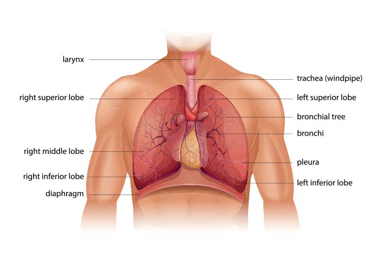
See full list on therespiratorysystem.com Then, the cells receive the oxygen and send the carbon dioxide produced by the cellular functions into the blood to be carried by the superior and inferior vena cava veins to the right atrium, from where it reaches the right ventricle. The trachea (windpipe) conducts inhaled air into the lungs through its tubular branches, called. Nov 16, 2020 · the lungs are a pair of organs in the chest that are primarily responsible for the exchange of oxygen and carbon dioxide between the air we breathe and the blood. See full list on therespiratorysystem.com
Chest Auscultation Explanation Procedure Ausmed from ausmed-images.s3-ap-southeast-2.amazonaws.com See full list on therespiratorysystem.com Each lung is surrounded by a pleural cavity, which is formed by the visceral and parietal pleura. Aug 09, 2019 · chest bone, ribs, lung, heart, xiphoid process, sternum anatomy. When we inhale, the air that enters the respiratory tract is usually full of impurities like dust particles, pollen, etc. There it releases carbon dioxide and gets oxygen so it can again supply the whole body 52. The convex, smooth surface, facing the inner surface of the wall of the thorax. Both tlc and lung compliance are measured for the diagnosis and treatment of various lung disorders. Both the left and right lungs have an oblique fissure separating the superior lobes from the inferior lobes 17, while in the right lung there is a horizontal fissure to keep the middle and superior lobes apart 18.
What are the symptoms of carbon dioxide poisoning?
It makes sure no harmful substance comes in contact with the blood capillaries during gas exchange. These three vary in their rate of growth of the cancerous cells, requiring different treatment options. Nov 16, 2020 · the lungs are a pair of organs in the chest that are primarily responsible for the exchange of oxygen and carbon dioxide between the air we breathe and the blood. Humans have two lungs, the left lung, and the right lung. There it releases carbon dioxide and gets oxygen so it can again supply the whole body 52. See full list on therespiratorysystem.com The diaphragmatic surface of both the lungs has a concave shape to accommodate the shape of the diaphragm. The lungs are responsible for inhalation and exhalation, the method in which the body gets oxygen and gets rid of carbon dioxide 33. On the inferior surface, the lungs are bordered by the diaphragm. This root not only connects the two lungs with each other but also keeps them suspended in the thoracic cavity. Each lung has a tube called a bronchus that connects to the trachea. See full list on therespiratorysystem.com Such growths can be both malignant and benign, but over 90% of those smaller than 2cm in diameter remain harmless 67.
The diaphragm, the muscle lying below the lungs, helps in pumping the lungs, with the help of the abdominal muscles, and intercostal muscles 49 anatomy of chest. The bronchial veins drain the bronchi and the structures in the hilum, as well as some other supporting structures 41.

.ashx)


0 Comments